Human Normal Peripheral Blood CD14+ Monocytes
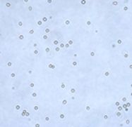
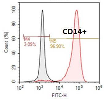
 Monocytes are often referred to as CD14+ monocytes, because of their expression of CD14. Monocytes make up between three to 8% of the leukocytes in the blood. Monocytes migrate from the bloodstream to other tissues and then differentiate into macrophages or dendritic cells. Monocytes and their macrophage and dendritic cell progeny serve three main functions, namely phagocytosis, antigen presentation, and cytokine production, in the immune system.
Our Human Normal Peripheral Blood CD14+ Monocytes are isolated from peripheral blood mononuclear cells by either positive or negative immunomagnetic selection, depending upon your needs. All peripheral blood is collected in acid-citrate-dextrose formula A (ACDA) by leukapheresis from fully consented IRB approved donors that are tested negative for HIV, HBV, and HCV.

Reference:
1. Loems Ziegler-Heitbrock and Thomas P. J. Hofer. Toward a refined definition of monocyte subsets. Front. Immunol., 04 February 2013
There are two major subsets of monocytes: CD14+CD16- monocytes and CD14+CD16+ monocytes. These subsets differ in terms of their phenotype, function, and gene expression profiles.
CD14+CD16- monocytes, also known as classical monocytes, are the most abundant subtype of monocytes, accounting for about 80-90% of all monocytes in the blood. They express high levels of CD14 and low levels of CD16 on their cell surface. CD14+CD16- monocytes are known for their ability to phagocytose (engulf and digest) bacteria and other microorganisms, and to secrete cytokines that initiate and regulate immune responses. They are also involved in tissue repair and angiogenesis.
CD14+CD16+ monocytes, also known as non-classical monocytes, are a minor subset of monocytes, accounting for about 5-10% of all monocytes in the blood. They express high levels of CD14 and high levels of CD16 on their cell surface. CD14+CD16+ monocytes are more pro-inflammatory than CD14+CD16- monocytes, and are involved in the regulation of T cell responses, as well as in the clearance of damaged cells and apoptotic bodies. They are also believed to play a role in the pathogenesis of inflammatory and autoimmune diseases.
In summary, CD14+CD16- monocytes are classical monocytes that are primarily involved in phagocytosis and cytokine secretion, while CD14+CD16+ monocytes are non-classical monocytes that are more pro-inflammatory and involved in the regulation of T cell responses and clearance of damaged cells.
Whole Blood Staining of Human Monocyte Subsets for Flow Cytometry
- Harvest human blood into a 13 ml Glass Whole Blood Tube with K3EDTA and mix the blood by gently inverting the tube several times.
- Transfer 100 μl whole blood into 5 ml polypropylene tubes and label the samples accordingly.
- Add 5 μl of each antibody into the blood sample and mix by gently tapping the tubes. (eg. anti-CD14, anti-CD16, etc.)
- Incubate the samples for 15 min in dark at room temperature.
- Add 2 ml 1x BD FACS Lysing Solution (10x solution diluted in ddH2O) in each sample.
- Vortex sample briefly three times, check for clarity and take to centrifuged within 2-3 min.
- Centrifuge 1,200 rpm for 7 min at room temperature.
- Remove supernatant and resuspend pellet in 200 μl 2% formaldehyde.
- Acquire the samples on BD LSR II flow cytometer.
- Analyze data using FlowJo.
Other Materials / Equipment Needed
- Human blood
- 10x BD FACS Lysing Solution (BD Biosciences, catalog number: 349202 )
- 20% formaldehyde (Tousimis, catalog number: 1008A )
- Phosphate buffered saline (PBS) (Life Technologies, Gibco®, catalog number: 20012-027 )
- Standard Bench-top Centrifuge
- 5 ml polypropylene tubes (BD Biosciences, Falcon®, catalog number: 352063 )
- BD LSR II flow cytometer
- Glass Whole Blood Tube with K3EDTA (BD Vacutainer®, catalog number: 366450 )


